

Mesenchymal stem cells and their exosomes sit on the edge of the next therapeutic wave for regeneration, inflammation control, and age-related disease…. but they are not miracle shots yet, and most of the proof still comes from animal and lab studies, plus a few small human trials. Used well, they may give safer, “cell-free” ways to calm inflammation, protect tissue, and push repair in skin, joints, heart, and brain.

Why this “next wave” matters
In clinic, people ask about “stem cell facials”, exosome serums, and IV drips almost every week now. There is a simple reason: more people live longer, and with that comes more chronic wounds, frailty, osteoarthritis, and memory loss.
Global data show the share of people over 60 may reach about 22% by 2050, which means hundreds of millions living with age-linked damage in skin, vessels, brain, and joints.
(User-supplied demographic data.)
Alzheimer’s disease alone is expected to affect roughly 3–5% of older adults, which keeps pressure on doctors and scientists to find better tools to protect tissue and slow decline.
(User-supplied epidemiology.)
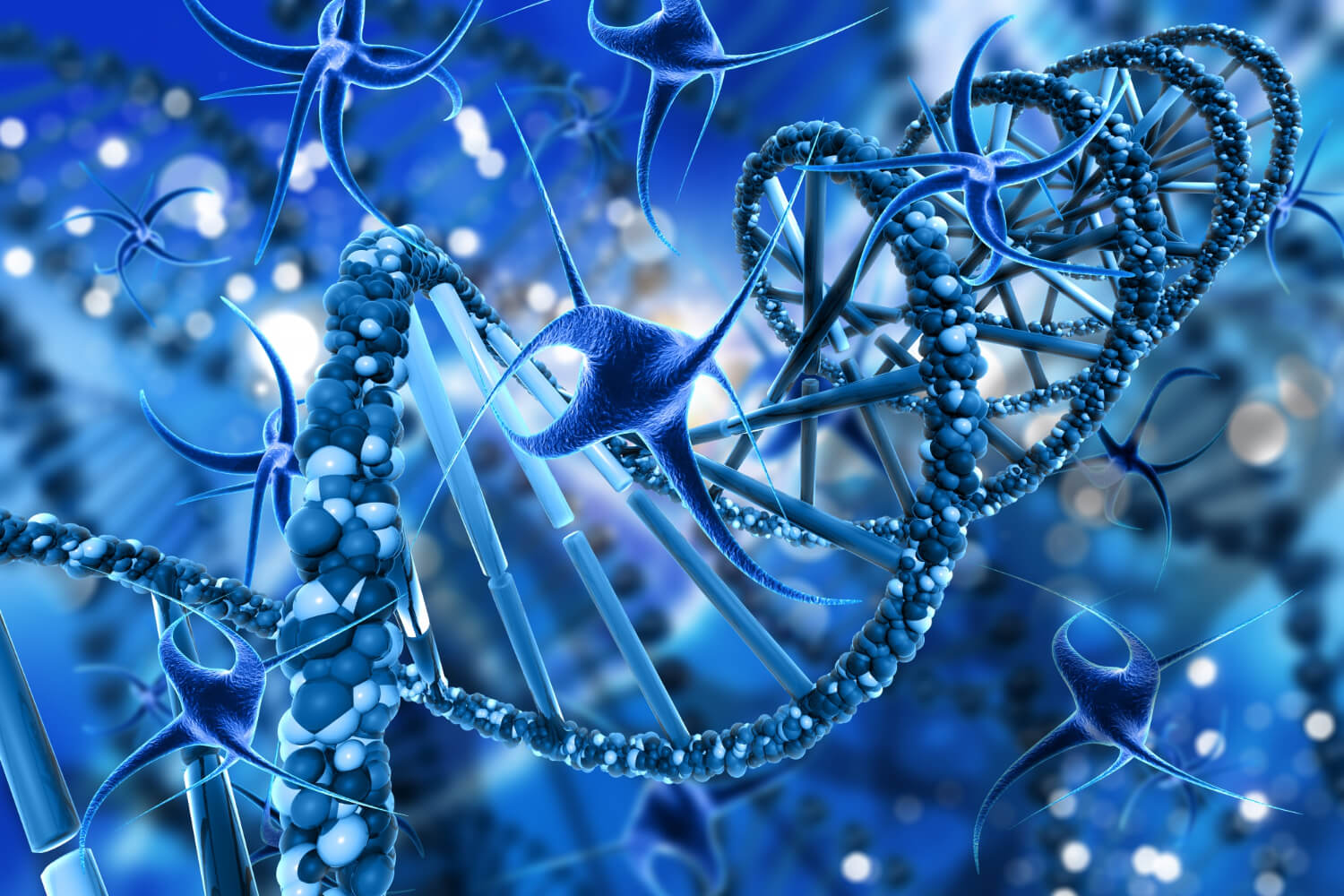
Mesenchymal stem cells (MSCs) and MSC-derived exosomes attract attention because they seem to talk to damaged tissue in a smart way: they carry proteins, lipids, and RNAs that shift immune cells, protect mitochondria, and nudge repair pathways rather than just “replacing” cells. The key is how they do this, and how far the science has really gone.
What MSCs and exosomes actually are
MSCs are multipotent stromal cells that can renew themselves and form bone, cartilage, and fat cells, and they live in bone marrow, fat, dental pulp, and umbilical cord tissue. In many injury models, MSC benefit comes less from them turning into new tissue and more from what they secrete into their surroundings.
Exosomes sit inside a broader family called extracellular vesicles (EVs). They are tiny spheres wrapped in a lipid membrane, usually about 30–150 nanometers across, formed inside multivesicular bodies and then released into blood, saliva, urine, and other fluids. Microvesicles tend to be larger (about 100–1000 nm), and apoptotic bodies even bigger (up to several micrometers), which helps labs sort these groups by size and origin.
MSCs release exosomes packed with:
- Signaling proteins
- MicroRNAs (miRNAs) and long non-coding RNAs
- Lipids and enzymes that tune inflammation and stress responses
Multiple reviews now list MSC-exosomes as key candidates for skin repair, chronic wound care, and age-related tissue damage, because they keep many helpful signals of the parent cells while avoiding some of the risks of whole-cell infusion.
Why exosomes may be safer than whole cells
When patients hear “stem cells”, they often picture cells floating through the body and building new tissue from scratch. The reality is less romantic and more practical…. and this is where exosomes shine.
Quick comparison: MSCs vs MSC-exosomes
| Feature | MSCs (whole cells) | MSC-derived exosomes |
|---|---|---|
| Size / structure | Living cells (≈10–20 µm), need to engraft or at least survive short-term | EVs 30–150 nm, lipid vesicles with no nucleus or cell division |
| Storage and logistics | Need careful freezing / thawing, lose function if handled poorly | More stable at low temps; easier to ship, store, and dose |
| Immune reaction and embolism | Can lodge in lungs or spleen; carry risk of immune reaction | Pass through microvessels more easily; lower immunogenicity |
| Tumor and ectopic growth risk | Theoretical risk of unwanted growth, fibrosis, or ectopic tissue | Do not divide, no direct tumor formation risk |
| Engineering options | Genetic change is complex and raises safety flags | Easier to load with drugs or specific miRNAs for targeted delivery |
Several groups show that MSC-exosomes can move through the microvasculature with less trapping in filter organs and lower chance of clots, because they are so small and lack the bulky cell body. They can also be frozen, stored, and thawed with less loss of function than living cells, which solves a big practical barrier for real-world use.
The other major plus: exosomes are easy to engineer. Labs attach targeting ligands on the surface or load the vesicles with specific RNAs, peptides, or drugs, creating what is basically a smart delivery capsule that homes to inflamed tissue and drops its payload there.
Skin healing and skin aging: where data look strongest
Skin is where most people see aging first, and it is also where MSC-exosomes have the richest preclinical data and the first controlled human trial signals.
Wound healing: three phases, many levers
A 2022 review of MSC-exosomes for cutaneous wound repair shows effects across all three classic phases of healing: inflammation, proliferation, and remodeling.
Inflammation phase
In animal wound and burn models, MSC-exosomes:
- Shift macrophages from the aggressive M1 state to the calmer M2 state through miRNAs such as miR-223 and let-7b, with changes in TLR4/NF-κB/STAT3/AKT signaling.
(User-supplied mechanistic data.) - Increase regulatory T cells and Th2 responses, which softens the inflammatory wave and prepares the wound bed for growth rather than chronic infection.
(User-supplied data.)
Proliferation phase
In vitro and in vivo, MSC-exosomes:
- Increase fibroblast movement and growth and improve capillary tube formation by activating AKT, ERK, and STAT3 pathways.
- Help keratinocytes survive oxidative stress through miRNA cargo such as miR-93-3p, which lowers pro-apoptotic APAF1, and activate Wnt/β-catenin to speed re-epithelialization in burn and incision models.
(User-supplied mechanistic data.)
Remodeling and scar control
Adipose-derived and umbilical-cord-derived MSC-exosomes:
- Raise matrix metalloproteinase-3, shift collagen from dense type I toward a higher type III ratio, and reduce myofibroblast build-up, which means finer, more flexible scars in animal models.
(User-supplied data.) - Deliver miRNAs such as miR-21, miR-23a, miR-125b, and miR-145 to fibroblasts and dial down TGF-β2/SMAD2 signaling, which is a main driver of hypertrophic scarring.
(User-supplied data.)
Taken together, preclinical work shows MSC-exosomes can calm “angry” immune cells, wake up sleepy fibroblasts and endothelial cells, and then fine-tune collagen so the final scar is thinner and softer.
Diabetic and ischemic wounds
Diabetic foot ulcers and ischemic leg wounds are two areas where the need is huge and standard care often fails.
In diabetic models, MSC-exosomes:
- Restore function of endothelial progenitor cells in high glucose, boost angiogenesis signals, and shrink ulcer area in animal feet.
(User-supplied data.) - Promote M2 macrophages, higher VEGF-A, and better re-epithelialization, while lowering stiff collagen I relative to collagen III, so healed skin is less tight.
(User-supplied data.)
In ischemic skin, exosomes loaded with TGF-β and adipose-MSC exosomes help fibroblasts move and close oxygen-starved wounds faster in animals.
(User-supplied data.)
Anti-aging and photoaging
Photoaging and “intrinsic” aging share common drivers: oxidative stress, DNA damage, and senescent cells that secrete inflammatory signals (the SASP). MSC-exosome work touches all of these:
- In human skin explants, umbilical-cord MSC exosomes enter the epidermis within about 18 hours and, after a few days of topical use, increase collagen I and elastin expression, hinting at actual matrix rebuilding.
(User-supplied data.) - Exosomes from iPSC-derived cells reverse senescence markers in UVB-damaged human dermal fibroblasts, raising collagen I and lowering SA-β-gal activity.
(User-supplied data.) - Adipose-MSC exosomes in photoaging models reduce ROS, DNA damage, and MMP-1, and increase type I collagen by tuning Nrf2 and TGF-β / Smad pathways.
A 2025 review on exosomes and skin aging notes that MSC-exosomes improve dermal thickness, collagen balance, and oxidative stress markers in several preclinical systems.
One real human trial: acne scars
For many readers, the big question is simple: “Has this helped real people yet?”
One randomized, double-blind, split-face trial used fractional CO₂ laser on atrophic acne scars and then applied adipose mesenchymal stem cell-derived exosome gel to one side of the face and control gel to the other.
Key points:
- 25 adults with atrophic acne scars completed three laser sessions plus gel.
- The exosome-treated side showed about 32.5% scar score reduction vs 19.9% on the control side, with less redness and shorter downtime.
Beyond skin: aging organs, brain, and heart
Oxidative stress and Nrf2
Several disease models show MSC-exosomes acting like tiny shields against oxidative stress:
- In UV-irradiated mouse skin and H₂O₂-stressed keratinocytes, MSC vesicles reduce ROS, DNA damage, abnormal calcium waves, and mitochondrial changes through Nrf2 activation.
- In intervertebral disc and osteoarthritis models, MSC-exosomes reduce NLRP3 inflammasome activation, ROS, and caspase-3/9 activity, easing cell death in cartilage and disc cells.
Nrf2 is a master switch for antioxidant genes such as SOD, GPx, and NQO1.
Wnt/β-catenin and tissue repair
A 2025 editorial from World Journal of Stem Cells highlights MSC-exosomes as strong activators of the Wnt/β-catenin pathway in hair follicles, kidney, lung, and heart.
In multiple models:
- BMSC exosomes increase β-catenin and TCF-4 while lowering pro-apoptotic Bax and cleaved caspase-3/9 in spinal cord injury and cardiac injury.
- Exosomes that deliver miR-29c-3p reduce BACE1, lower amyloid-β (Aβ1-42), and reduce IL-1β, TNF-α, and IL-6 in Alzheimer-type models.
SIRT1 and chronic inflammation
Sirtuin-1 (SIRT1) is a key longevity-linked enzyme. In myocardial infarction models, MSC exosomes delivering lncRNA KLF3-AS1 regulate the miR-138-5p/SIRT1 axis, cutting cardiomyocyte inflammation and cell death, with better heart function in rats.
(User-supplied data.)
Amniotic MSCs engineered to overexpress SIRT1 reduce arthritis scores and inflammatory cytokines, while increasing Treg cells and protecting cartilage in mice.
(User-supplied data.)
In acute UV damage, hUCMSC exosomes increase SIRT1 in HaCaT keratinocytes through transfer of 14-3-3ζ protein and stronger autophagy, which lowers oxidative injury from UV and H₂O₂.
(User-supplied data.)
Safety, regulation, and what this means for patients
Where the evidence stands
- Most data for MSC-exosomes are still preclinical.
- Human data come mainly from MSC cell trials.
- Controlled exosome-only trials are young.
Manufacturing and quality control
- Pure exosome isolation is almost impossible.
- GMP standards are required.
- Thrombosis risk must be monitored.
- Potency testing is still under debate.
How to think about this as a reader or patient
- Treat MSCs and exosomes as experimental tools.
- Ask for peer-reviewed human data.
- Be cautious with IV “exosome drips” outside trials.
Aging is real. So is the hope that smarter, cell-free biologics like MSC-exosomes will help people heal better and stay functional longer.
Why some patients travel to Mexico for therapy
I see many patients from the United States who quietly tell me the same thing…. “Doctor, I just can’t afford this care at home.”
That is one big reason more people now travel to Mexico for stem cell and exosome-based treatments. Clinics such as R3 Stem Cell Mexico offer mesenchymal stem cell and exosome procedures at a lower price point than many U.S. centers.
Several of my patients who went there share stories of kind staff, clear communication in English, and structured follow-up after treatment.
Cross-border travel for regenerative procedures has grown as more centers in North America and Latin America offer MSC and extracellular-vesicle-based care.


